 Multiple Choice Questions
Electron microscopes have much higher resolution than any type of light microscope because
of their higher maginification.
the lenses used are of much higher quality
of very short wave length of electrons.
the images are viewed on screen rather than directly using an eye-piece or occular lens.
A bioinformatics tool used to find the sequence similarity in the subunits of hemoglobin is
FASTA
BLAST
HUMMER
PSI:PLOT
Which one of the following molecular marker types uses combination of both restriction enzyme and PCR techniques?
SSR
AFLP
SNP
RAPD
An ant moving straight, upon encountering an obstacle, may turn either right or left and continue moving. To test the hypothesis that the direction chosen by the ant is random, the most appropriate statistical test is
Student's t-test
χ2-test of independence
χ2 -test of goodness of fit.
correlation test
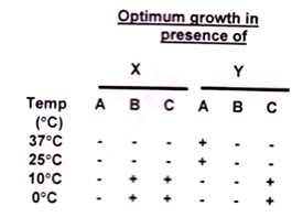
To test whether bacteria with enhanced toluene degradative abilities could be created for a cold environment, a TOL (toluene-degrading) plasmid from a mesophilic bacterial strain was transferred by conjugation into a facultative psychrophile. The psychrophile was able to degrade salicylate (SAL) but not toluene. The recombinant strain carried the introduced TOL plasmid and its own SAL plasmid. The results are as follows:

Identify A, B, C, X, and Y.
A - mesophile; B - psychrophile; C - Transformant; X - Toluene; Y - Salicylate.
A - mesophile; B - psychrophile; C - transformant; X - Salicylate; Y - Toluene
A - psychrophile; B - transformant; C - mesophile; X - Salicylate; Y - Toluene.
A - transformant; B - psychrophile; C - mesophile; X - Toluene; Y - Salicylate.
A small fraction of clear cellular lysate was run on an isoelectric focusing gel (IEF) to purify a particular protein, which showed a number of sharp bands corresponding to different pI values. The protein of interest has a pI of 5.2. Therefore, the band corresponding to pI 5.2 was cut, eluted with appropriate buffer and subjected to SDS-PAGE, which showed 3 distinct bands. Which one of the following inferences CANNOT be drawn from the above observations?
Several different proteins having the same pI may be present at the single band on IEF gel.
SDS-PAGE showed 3 distinct bands that may represent molecular mass of different proteins.
The protein of interest may be composed of 3 subunits.
As the IEF gel showed a distinct band corresponding to pI 5.2, which is the pI of the protein of interest, the protein is composed of a single subunit.
Chromosome organization becomes clearer from a series of biochemical, electron-microscopic and X-ray crystallographic studies. When interphase chromatin is isolated in low salt buffer on string organization is seen. Interphase chromatin directly observed under EM shows 30 nm fibre. When histones are depleted from metaphase chromosome and visualized under EM, it shows a huge number of very large loops associated with scaffold.
Following interpretations can be made from these:
A. 11nm fibre is formed when nucleosomes are brought closer by scaffold.
B. 30nm interphase chromatin is formed by zig-zag organization of nucleosomes of 11 nm fibre.
C. 30nm fibre makes a solenoid packing to form the metaphase chromosome.
D. 30nm fibre gets organized into loops due to SARs getting associated with scaffold proteins and coming closer.
The correct combination of interpretations is
A and D
A and C
A and B
B and D
Cytochrome-c has only one tryptophan residue (W) which is buried. The protein in cacodylate buffer (pH 6.0) is excited at 280 nm, and its emission spectrum measured in the range of 300-450 nm. The same measurement was repeated on the protein in the buffer containing 6M guanidine hydrochloride. It was observed that there is an increase in the intensity of the emission spectrum of the guanidine hydrochloride treated cytochrome-c. The most probable reason for this increase is:
W is near hydrophobic patch present in the unfolded protein.
W is near the heme in the native protein.
W is near the carboxylate amino acid side chains in the native protein.
W is in a polar pocket in the native protein.

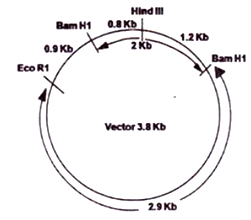
The above diagram represents a 2Kb insert successfully introduced in between two BamHI sites of a 3.8 Kb vector in desired orientation. The HindIII site on the insert and EcoRI site on the vector is also indicated. If the insert was introduced in the opposite orientation, which one of the following statements is INCORRECT?
Digestion with EcoRI will linearize the 5.8 Kb plasmid
Digestion with BamHI will yield one 3.8 and one 2.0 Kb fragment.
Double digestion with EcoRI and HindIII will produce 1.7 Kb and 4.1 Kb fragments.
Double digestion with EcoRI and HindIII will produce 2.1 Kb and 3.7 Kb fragments.
D.
Double digestion with EcoRI and HindIII will produce 2.1 Kb and 3.7 Kb fragments.
Among the given statements, the statement that in incorrect for the diagram is that double digestion with EcoRI and HindIII will produce 2.1 Kb and 3.7 Kb fragments.
The hydrogen atoms in the δ (delta) methylene group of lysine will give the following splitting pattern in the 1HNMR spectra of lysine.
Triplet of triplets
Quintet
Double of triplets
Triplet of a doublet
